English translation updated . This article reflects the source material at its original publication date. Read the Chinese source.
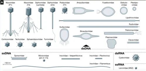
Many tools in modern molecular biology grew out of research on phages. Studies of λ phage informed numerous techniques. T7 phage, shown in Figure A, provided the DNA polymerase from which Sequenase was developed, while the T7 RNA polymerase system became an important tool for heterologous protein expression and in-vitro transcription.
The Microviridae phage φX174 was the first genome sequenced by Fred Sanger. Arthur Kornberg used it as a model to demonstrate that DNA synthesized with purified enzymes could be biologically active. Craig Venter’s work subsequently demonstrated biological activity from a genome assembled using synthetic oligonucleotides.
MS2, described in the source under the historical family Leviviridae, infects Escherichia coli and encodes only four proteins. The interaction between its coat protein and an RNA hairpin derived from its operator sequence became the basis of an influential system for imaging specific RNAs in living eukaryotic cells.
Four prophages, φNM1–4, identified in Staphylococcus aureus prompted research into phage-based payload delivery. Components from φNM1 were used to produce capsids carrying a CRISPR/Cas9 system targeting S. aureus. The cited work investigated killing antibiotic-resistant staphylococci. The original Chinese text incorrectly calls MRSA a virus; MRSA is a bacterium.
Filamentous phages encompass diverse structures and lifestyles. The source describes particles ranging from approximately 800 nm to 4 μm in length, including particles longer than the bacterial cells they infect. Taxonomic family names in this historical article reflect the terminology used in its 2019 source.
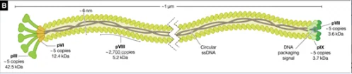
Figure B. Schematic of E. coli Ff phages, including overall structure and structural-protein copy numbers.
Ff filamentous phages
Much of our understanding of filamentous phage biology comes from the closely related E. coli phages M13, fd and f1, collectively termed Ff phages. They share approximately 98% DNA sequence identity and similar replication mechanisms. They were independently isolated from sewage samples in the early 1960s.
A common feature of Ff and many other filamentous phages is that they do not lyse their hosts. In bacterial lawns, infection produces zones of reduced growth rather than the clear plaques characteristic of strongly lytic phages. Ff phages are valuable model systems, but they do not represent the full diversity of filamentous phages.
M13 provided one of the early cloning vectors used for DNA sequencing. Ff phages also underpin phage display, which links protein–protein interaction screening with the selection and maturation of protein, peptide, antigen and antibody libraries.
Several properties make them useful:
- Particle assembly does not lyse the E. coli host, allowing continued release during culture. The source reports titres reaching 10¹³ particles per millilitre.
- Coat proteins can be genetically fused to other proteins, displaying foreign sequences on the particle surface.
- The small genome is readily modified, and each particle packages the genome encoding its displayed protein. This physical link between phenotype and genotype allows both a selected molecule and its corresponding gene to be recovered and improved through repeated selection.
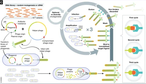
Figure C. Phage display.
Phage display and selection
In the system described, a natural or synthetic DNA library is inserted between the sequences encoding a signal peptide and mature pIII in a vector containing an Ff replication origin, a plasmid replication origin and a selectable marker. The vector is introduced into E. coli, while a helper system supplies the components needed for phage replication and assembly. The resulting particles display a proportion of modified pIII proteins containing the inserted sequence.
The library undergoes repeated rounds of selection, or “panning.” Particles are exposed to an immobilized ligand or target; those with suitable binding properties remain while nonbinding particles are removed. Bound particles are recovered and amplified for subsequent selection rounds. Repeated selection can enrich peptides with improved affinity.
The 2018 Nobel Prize in Chemistry recognized work on directed evolution, including Gregory Winter and George Smith’s development of phage display for peptides and antibodies, alongside work on enzyme evolution.
Source
Hay ID, Lithgow T. Filamentous phages: masters of a microbial sharing economy. EMBO Reports. 2019;20(6):e47427. Published online 5 April 2019. doi:10.15252/embr.201847427.