English translation updated . This article reflects the source material at its original publication date. Read the Chinese source.
Carrier-mediated intracellular phage delivery
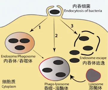
Research on intracellular phage delivery often uses carriers to improve uptake. Phages are macromolecular particles and cannot cross cell membranes by passive diffusion. They enter through processes such as macropinocytosis, phagocytosis and transcytosis. See the original related article on intracellular infections.
Encapsulating phages in, or attaching them to, a carrier can act as a “Trojan horse.” Broxmeyer and colleagues described delivery using nonpathogenic Mycobacterium smegmatis infected with phage TM4. Neither the bacterium nor TM4 alone reduced intracellular M. tuberculosis or M. avium. When phage-infected M. smegmatis was taken up by cells, however, it colocalized with M. avium in vacuoles. The reported intracellular bacterial load fell approximately 100-fold after 48 hours [2]. Danelishvili and colleagues subsequently reported reduced splenic bacterial loads in mice with disseminated M. avium infection treated with TM4-infected M. smegmatis. Although innovative, using live bacterial carriers in patients presents risks.
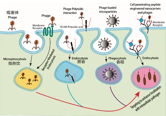
Figure 2. Cellular uptake of phages [1].
1. Liposomes
Lipid nanovesicles made from natural or synthetic lipids are versatile delivery systems. They self-assemble into closed bilayer spheres whose interior can protect sensitive cargo. The studies discussed in the source describe compatibility between liposomes and phages during and after encapsulation. Potential benefits include delivery to target sites and into cells, and protection from low pH, reticuloendothelial clearance and neutralizing antibodies.
In one experiment, approximately 100 nm cationic liposomes prepared by lipid-film hydration encapsulated KPO1K2 phages with an average size of approximately 50 nm. This supported entry into Klebsiella pneumoniae-infected macrophages. The reported killing of intracellular bacteria was 94.6% for liposomal phages, compared with 21% for unencapsulated phages after 24 hours. Encapsulation also protected phages against neutralizing antibodies in the reported experiments [3].
Surface modification with dextran, chitosan, alginate or synthetic polymers such as polyethylene glycol or polyvinyl alcohol can alter liposome properties. Polyethylene glycol can provide a hydrophilic surface layer. The source also describes reports that conjugating the tetrapeptide tuftsin to liposomes increases uptake by mononuclear phagocytes, suggesting a possible route for delivering encapsulated phages against intracellular bacteria. This remains a research proposition.
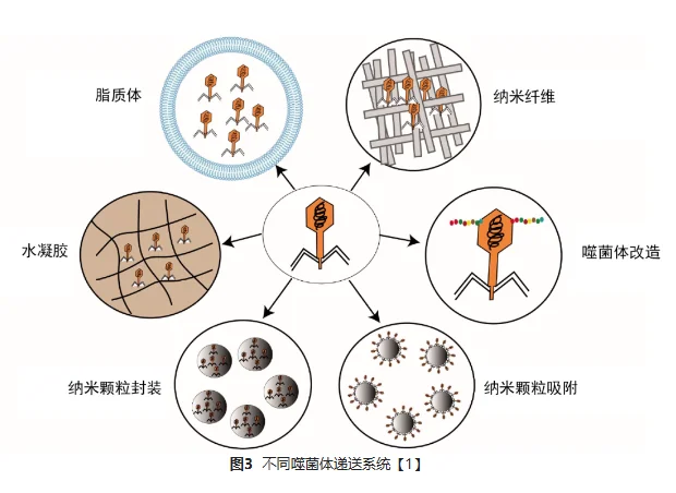
2. Nanomaterials
Nanomaterials provide another approach to phage delivery. Fulgione and colleagues reported that biomimetic hydroxyapatite (HA) nanocrystals could deliver Salmonella phage SR φ1 into cells [4]. HepG2 cells infected using a bacterial suspension of 10⁸ CFU/mL were treated for 24 hours with equal quantities of free phages or HA-loaded phages. The source reports approximately 10⁵ CFU/mL following HA–SR φ1 treatment, compared with 10⁸ CFU/mL for free phages. Improved stability at pH 4.0 suggested potential value in acidic compartments such as phagolysosomes.
Electrospinning can produce nanofibres with controlled surface morphology, a high surface-area-to-volume ratio, flexibility, functionalizable surfaces and useful mechanical properties. Incorporating bioactive cargo can protect it before release. Responsive fibres could release phages in response to changes in pH, temperature or other stimuli.
The source discusses Eudragit® methacrylate polymers as pH-responsive encapsulation materials. It lists a dissolution threshold of approximately pH 6 for Eudragit L100 and pH 7 for S100. It then proposes that L100 would retain phages in a neutral extracellular environment and release them as intracellular pH falls. Those two statements are inconsistent with the dissolution thresholds given in the same source. The proposed acid-triggered mechanism requires checking against the original study and is not presented here as an established delivery mechanism. The general research objective is to protect phages during transport and release them at an appropriate site.
3. Small molecules and peptides
Peptides or small molecules attached to a carrier or phage surface can influence cellular uptake and targeting. Hussain and colleagues conjugated vancomycin-loaded nanoparticles to the cyclic nine-amino-acid peptide CARGGLKSC (CARG). CARG targeted bacterial surface components and accumulated in staphylococcal infections of the lung and skin. The source reports complete recovery and long-term survival in the treated infected mice [5]. This example concerns antibiotic nanoparticles, not a clinical phage treatment.
Engineered phages displaying peptides such as cell-penetrating or integrin-binding sequences have also been investigated to promote cellular uptake and organelle targeting.
The archived article contains reference numbers [1]–[5] but no corresponding bibliography. These citations are retained for traceability; their full bibliographic details require confirmation.